Hasiera » Platforms » Achucarro Imaging Platform » Structured Illumination

Imaging Platform | Structured Illumination

Carl Zeiss ApoTome2

The Carl Zeiss ApoTome2 microscope can create optical sections of fluorescent samples (free of scattered light). The structured illumination of this equipment projects a grid structure into the focal plane of the specimen, and then moves it into three positions using a scanning mechanism..

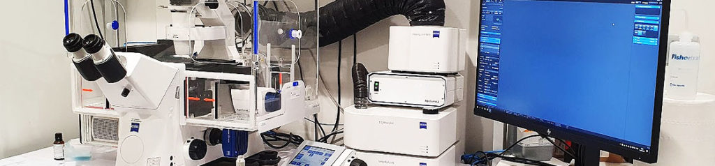
  • Automatic grid selection for optimum contrast with each objective (2.5 – Dry 10 – Dry 20 – Dry 40 – Dry 40 – Oil 65 – Oil)
  • Freely selectable grid, depending on the specimen
  • Combination of multiple contrast techniques: Fluorescence channels (DAPI, GFP, DsRed), brightfield, darkfield, DIC, polarized light.
  • Live Cell Imaging and time lapse (with CO2 and temperature control system)
  • Up to three images per second (depending on the exposure time). Doubling of frame rate with “burst mode” (in Live Cell Imaging)
REQUEST ACCESS

Please check the user policy and then access the access request form: