Home » News » New Imaging Method Maps Tiny Protein Clumps Linked to Parkinson’s Disease
New Imaging Method Maps Tiny Protein Clumps Linked to Parkinson’s Disease
16Oct2025
The international group of scientists developed a powerful optical technique that can spot and map the earliest, most toxic forms of the protein α-synuclein in brain tissue.
The discovery could help reveal how Parkinson’s disease develops and pave the way for better diagnostics and treatments.
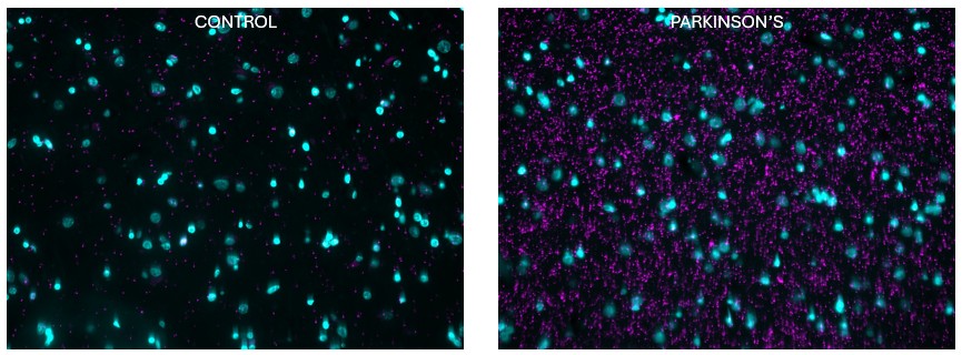
In this paper recently published in Nature Biomedical Engineering, the authors report a new optical method that can find and map tiny clumps of the protein α-synuclein (often written “Asyn”) in human brain tissue — the small, soluble clumps called oligomers that many scientists think trigger Parkinson’s disease.
By using very sensitive single-molecule fluorescence imaging, the team made large-scale, nanoscale maps showing where these oligomers sit, how many there are, and how big they are in post-mortem Parkinson’s brains. That matters because most previous studies could only see large fibrils or small fields at a time; this technique lets researchers look across whole tissue regions and count the potentially toxic species, which could help track disease processes and guide future diagnostic tests or treatments.
One of the principal investigators involved in the study is the Ikerbasque and Ramón y Cajal researchers in ACHUCARRO and EHU, Dr Nora Bengoa-Vergniory that commented “it’s an important step to clarify the biology of Parkinson’s which could also open new avenues for diagnosis and treatment”.