
 Main technical specifications
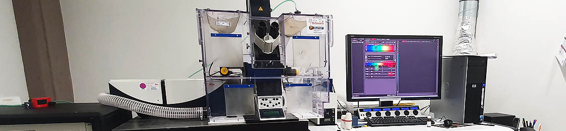
- Inverted Leica DMI 6000B microscope.
- Objectives: 10x Air, 20x Air, 40x Oil, 63x Oil, 100x Oil (g-STED).
- Galvanometric Z-stage and motorized XY-stage for multiposition imaging (mosaic, time lapse).
- White Light Laser (470-670 nm tunable excitation with 1nm interval).
- Argon Laser (458, 476, 488, 496, 514 excitation lines).
- Ultraviolet Laser (405 nm excitation line, AOTF Flexible)
- STED CW unit (592 depletion laser for super-resolution and 100x objective)
- Galvanometric Scanner (up to 1400 Hz) and Resonant Scanner (8000 Hz) for fast scanning.
- High-sensitive Hybrid detectors (x2) and standard Photomultiplier tubes (PMT) (x2): 4 channel simultaneous acquisition with freely adjustable spectral detection.
- Environmental control chamber (temperature, CO2, humidity) for in vivo imaging.
 Main technical specifications
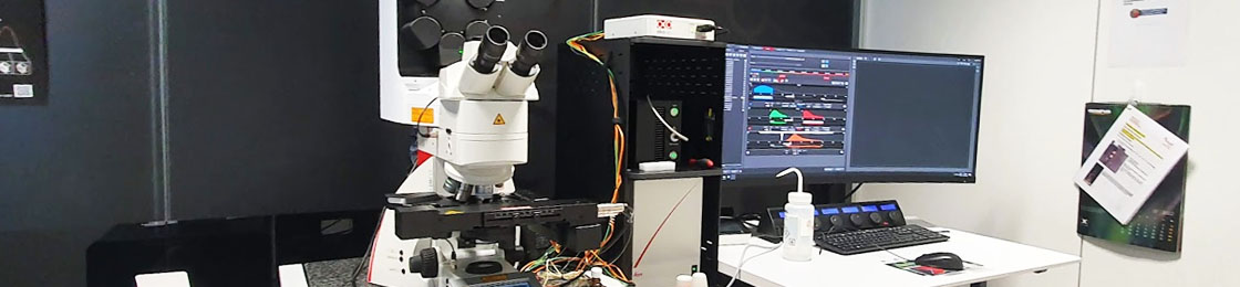
- Objectives: HC PL APO 10x/0.40 Air CS2, HC PL APO 20x/0.75 Air CS2, PL APO 40x/1.30 Oil CS2, PL APO 63x/1.40 Oil CS2, PL APO 40x/1.10 Water CS2, HC APO L 40x/0.80 W U-V-I diping
- Galvanometric Z-stage and motorized XY-stage for multiposition imaging (mosaic, time lapse).
- Second generation White Light Laser (WLL2).
- Ultraviolet Laser (405 nm excitation line, AOTF Flexible)
- Second generation Hybrid detectors (x3)
- Supercontinuum Fiber Laser: Excitation range between 485 – 685 nm. in 1nm steps. Pulsed laser. Pulse frequency: 80MHz (compatible with lifetime measurement applications for processing). Laser lines controlled by AOTF and AOBS that allow to vary power and the simultaneous use of up to 8 wavelengths simultaneously.
- TauSense module
- LIGHTNING super resolution module
The Femto-2D microscope allows fluorescence imaging of neurons in living brain slices and in vivo. In addition to imaging live cell morphologies, it can be used for calcium imaging and other functional fluorescence microscopy approaches. The setup is based on a tunable femtosecond laser equipped with a DeepSee module to compensate pulse dispersion and facilitate imaging deeper inside tissue. GaAsP and PMT detectors, respectively, allows for dual color imaging. The upright microscope is additionally fitted with electrophysiology equipment for two channel field or whole-cell recordings, as well as an LED epifluorescence module.
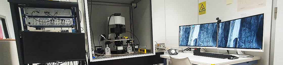
Main technical specifications
- Can be configured for in vivo or in vitro studies
- Two channel fluorescence detection
- High precision galvanometric scanning
- MaiTai 690 – 1040 nm tunable femtosecond excitation laser
- DeepSee pulse dispersion compensation
- Fully equipped for patch-clamp electrophysiology
- Two color LED epifluorescence
 Main technical specifications
- Automatic grid selection for optimum contrast with each objective (2.5 – Dry 10 – Dry 20 – Dry 40 – Dry 40 – Oil 65 – Oil)
- Freely selectable grid, depending on the specimen
- Combination of multiple contrast techniques: Fluorescence channels (DAPI, GFP, DsRed), brightfield, darkfield, DIC, polarized light.
- Live Cell Imaging and time lapse (with CO2 and temperature control system)
- Up to three images per second (depending on the exposure time). Doubling of frame rate with “burst mode” (in Live Cell Imaging)
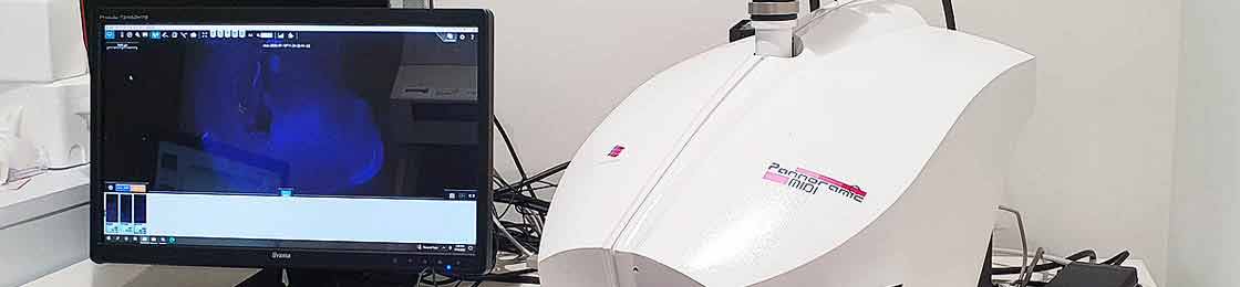
Automated digital slide scanner (virtual microscopy equipment) by 3DHistech, our Pannoramic MIDI II has the following specifications:
Main technical specifications- Scan method: area scanning with autofocus
- Z-stack imaging feature
- 12 slide capacity with automatic sample recognition and digitization of sample detection
- Light sources: Wideband Multichannel RGB LED light source for brightfield operation
- Advanced fluorescent light source: Lumencor SPECTRA 6 solid state light engine
- Filters set: DAPI, FITC (A488), TRITC, TexasRed (A594), Cy5
- Magnification: Plan Apochromat 20x/0.8 (dry) Plan Apochromat 40x/0.95 (dry)
- Image digitization: 4.2 MP Scientific sCMOS digital imaging camera (for brightfield and fluorescence)
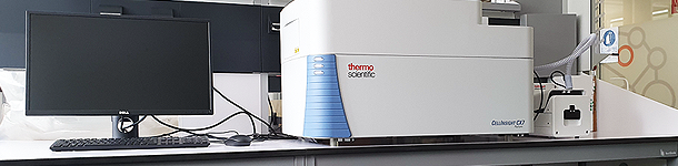 We have one ThermoFisher CellInsight CX7 High-Content Screening Platform that enables a combination of cell biology with automated high resolution microscopy and robotic handling to extract the information from samples using several imaging modes. It covers the entire fluorescence spectrum and captures images selecting either widefield or confocal optics for any channel. The platform is also combined with an Onstage Incubator that allows precise control of temperature, humidity and CO2 levels in order to observe and measure biological activity and changes over time.
Main technical specifications
We have one ThermoFisher CellInsight CX7 High-Content Screening Platform that enables a combination of cell biology with automated high resolution microscopy and robotic handling to extract the information from samples using several imaging modes. It covers the entire fluorescence spectrum and captures images selecting either widefield or confocal optics for any channel. The platform is also combined with an Onstage Incubator that allows precise control of temperature, humidity and CO2 levels in order to observe and measure biological activity and changes over time.
Main technical specifications
- 7-channel solid-state LED light engine
- Photometrics X1 camera Widefield
- 7-channel fluorescent imaging, 5-channel brightfield imaging, 7-channel confocal imaging
- 3-position objective (range from 2x–40x, low and high NA)
- Software and laser-based autofocus for consistent scan times
- Configured for fully automated plate handling and scanning
- HCS Studio software for integrated data collection and analysis
ULTRAMICROTOME
We have one PC controlled ultramicrotome by RMC Boeckeler, with an integrated touch screen control monitor, that provides good training capabilities. This equipment can take ultra-thin sections – to be then studied in the Electronic Microscopy Laboratory, within the Advanced Research Facilities (SGIker) of the University, on the campus -.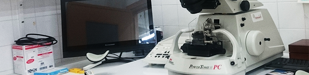 Main technical specifications
- Auto thin sectioning from 1nm and Auto thick sectioning to 15µm
- Cutting speed range from 0.1 – 100mm/sec
- Specimen auto feed 200µm
- High precision manually operated knife stage with 30mm lateral and 12mm forward motion
- Upper knife stage with 360° rotation and knife clearance angle adjustment from -2° to +15°.
- Accepts glass knives up to 12mm wide, triangular tungsten carbide knives,…
- Cutting zone set by its control system that can be adjusted at any time.
- Numeric & digital displays of number of sections cut, total specimen feed advanced, section thickness, cutting speed and specimen feed remaining
- Stemi 2000 stereomicroscope with 7.7:1 zoom range, 6.5X to 50X magnification with 10X wide field (23mm) eyepieces