Imaging Platform | Structured Illumination
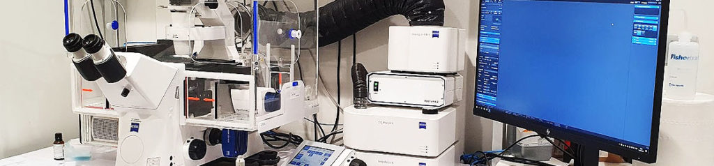
Carl Zeiss ApoTome2
The Carl Zeiss ApoTome2 microscope can create optical sections of fluorescent samples (free of scattered light). The structured illumination of this equipment projects a grid structure into the focal plane of the specimen, and then moves it into three positions using a scanning mechanism..

- Automatic grid selection for optimum contrast with each objective (2.5 – Dry 10 – Dry 20 – Dry 40 – Dry 40 – Oil 65 – Oil)
- Freely selectable grid, depending on the specimen
- Combination of multiple contrast techniques: Fluorescence channels (DAPI, GFP, DsRed), brightfield, darkfield, DIC, polarized light.
- Live Cell Imaging and time lapse (with CO2 and temperature control system)
- Up to three images per second (depending on the exposure time). Doubling of frame rate with “burst mode” (in Live Cell Imaging)